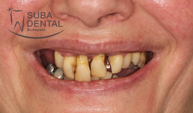
Completely edentulous jawbone and a bar denture retained by 4 implants (37) (Case presentation)
Our female patient aged 56 presented to Suba Dentál’s clinic with the following complaints:
- Her teeth were very loose.
- She had bad breath (halitosis).
- She desired to have an aesthetic smile.
- She desired a stable, palateless solution possibly by means of implants

Before
 Aftera
First treatment (3 workdays): tooth extractions:
All teeth had to be removed due to a severe chronic periodontitis and dental foci at the first treatment. Had her periodontal disease been treated several years earlier and had she undergone professional tartar removal at least twice a year and had her teeth been treated by a periodontist her gums, teeth and the jawbone would never have sunk into such a state of disrepair. Once the teeth are extracted it is equally important to remove the inflamed granulation tissues, pus pockets and deep periodontal pockets in order to ensure proper healing for implants to be installed. Simultaneously with the elimination of dental foci a provisional denture was prepared to provide teeth for the 3-month healing period. Gums at the tooth extraction sites were sealed with sutures which had to be removed after 10 days of healing. Most of the time resorbable sutures are used nowadays, which come off on their own after 3 to 4 weeks.
Important notice!
- The provisional denture, oral cavity and the tongue should be thoroughly cleaned with a tooth brush and toothpaste following each meal.
- If possible, the provisional denture should only be worn at your workplace, and it should be removed at home in the interest of uncomplicated healing.
- A few weeks on, the inflammation and swelling resolves causing the provisional denture to become loose and tilt, which can be remedied by a reline or denture adhesive.
- The dentures should be removed overnight and stored in a clean condition, enabling the fresh wound to “breathe” and heal properly.

The first panoramic radiograph presented by our patient at the first consultation It readily appears that the bone around the teeth has almost completely resorbed and what we are facing here is severe periodontal disease.

Follow-up panoramic radiograph recorded after the first treatment. The teeth and the inflamed tissues were removed.
A few photographs capturing the initial condition:


Second treatment – 3 workdays, tooth implantation
Following 3 months’ healing after tooth extractions a CBCT (Cone beam computed tomography) was recorded in order to examine residual bone mass in 3 dimensions prior to the implant surgery. Based on the scan we were able to assign the precise location and size of the implants. 4 implants were installed in the lower and upper arch each, which would accommodate a bar to ensure exceptional stability for the removable denture. The stability of the lower implants exceeded 30 Ncm warranting the placement of healing abutments (gingiva formers). The stability of the upper implants did not attain 30 Ncm as a result of which cover screws were placed in the implants. Following surgery the gums were sealed with sutures which had to be removed 10 days after the intervention. The provisional dentures the patient began to wear after the removal of her teeth were relined so she could wear them for the remaining 6 months of the healing period.

CBCT scans of the lower right region. The lower middle image demonstrates a bone height of 12 mm.

CBCT scan of the upper right region. Bone height only ensures the installation of an implant of length not exceeding 10 mm.

Follow-up panoramic radiograph following installation of the implants. It can be readily seen that the stability of the lower implants allowed for the installation of healing abutments (gingiva formers) whereas the upper ones were closed with cover screws due to the lesser stability of the upper arch.
Third treatment, 1 workday, the exposure of implants
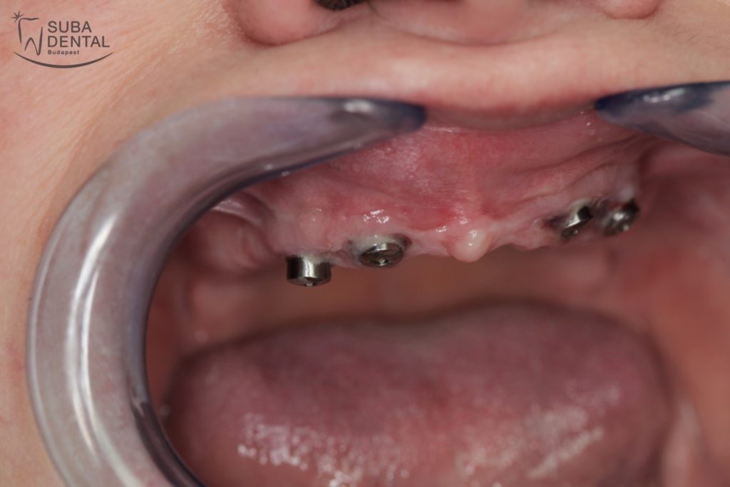
Prior to installing the permanent replacement our patient underwent a minor surgical intervention in which the upper implants were exposed individually and healing abutments were placed therein. The healing abutments would shape the gums in the weeks ahead to give the permanent abutment and tooth replacement a perfect gum contour. The provisional denture obviously required to be relined due to the change.

The maxilla following implantation and healing. Cover screws are placed in the implant sealing invisibly inside the bone underneath the gums.

Gingiva formers (healing abutments) placed inside the implants of the upper arch. The healing abutment (gingiva former) protrudes from the gums and shapes them appropriately in order for the replacement tooth to sit perfectly in the implant.
Fourth treatment – 10 workdays, the preparation of the permanent tooth replacements
Following the 6-month healing period it was time we set about fabricating the permanent replacement. Precision impressions were taken of the implants with closed tray impression copings.

Healing abutments in the implants installed in the mandible.

The gums around the implants of the mandible perfectly shaped by the healing abutments (gingiva formers).
Following impression taking several test fittings had to be performed while the dentures were being prepared by the dental technician.

Follow-up panoramic radiograph with the closed-tray impression copings placed inside the upper implants.

Photograph of the closed-tray impression copings placed inside the lower implants prior to impression taking.
During the bar test fitting the accuracy of the impressions and the trueness of the bar supporting the tooth replacement are verified. All tooth replacements must seamlessly fit the master cast.

The lower bar on the master cast

The lower bar on the master cast with a matching screw driver

The lower bar with its screws connecting to the implants installed in the bone removed from the master cast.
During the tooth test fitting the bite (function) and aesthetics (colour and shape of the teeth, the middle line and the smile line) are checked.

Tooth test fitting with test teeth embedded in wax.
Completion, handover: when everything is perfect the dentures are ready to be finished.

The finished bar-retained denture on a gypsum cast.

The metal framework of the bar-retained denture designed for the mandible with built-in Dolder bar retention.
Following delivery of the new tooth replacement the patient returns for follow-ups in which the condition of the gums is checked (for any cracks) and the bite (occlusion) is precisely adjusted.
The patient was satisfied on completion of the treatment. She had to get accustomed to her mouth being “full of teeth” again. With teeth filling her oral cavity again her face has become youthful again, the lips and the soft skin around mouth “plumped out”. Our patient is at ease to smile and mingle with and talk to people again.
IMPORTANT NOTICE!
- Implant tooth replacements require as much, if not more attention, care and follow-up as natural tooth-borne tooth replacements. There is close relationship between oral hygiene, the quantity and quality of tartar and the long-term success of the implants.
- It is a common misconception that tartar cannot cling to tooth replacements/implants. Implant borne tooth replacements are as prone to tartar-build up as natural teeth are.
- Following the completion of removable permanent dentures a reline is required every 2 to 3 years. In the absence of root canals the jawbone is in a constant and piecemeal process of deterioration. Unfortunately this is a normal physiological process in which the bone slowly resorbs, which is adversely affected by the pressure exerted by the removable denture.
- Out-of-true, waggling dentures accelerate bone resorption, and therefore necessitate annual follow-ups where the dentist performs a bite check and establishes the extent of waggling to determine whether a reline is necessary. If the patient fails to show up at the follow-ups, and no bite correction and reline are performed, the loose, out-of-true dentures can exert forces severely damaging the implants.
- Traumatic occlusion, tartar and an out-of-true denture may cause periimplantitis, as a result of which the bone resorbs, which is marked by the thread of the implant becoming visible. Tartar and bacteria easily adhere to the ragged surface of this “protruding” implant which may entail the rejection of the implant.
- Warranty is therefore subject to annual follow-ups, tartar removal and the performance of panoramic radiography.

Follow-up panoramic radiograph with the finished bars on completion of the treatment.
The finished bars in the mouth:


The finished bar-retained dentures in the mouth

Before

After


